Home

arabulucu yalnız Yan ürün koedam kappa Siber alan bariyer ayrılık

Scott Koedam - Area Manager - Dakota Matting and Environmental Solutions |  LinkedIn
Scott Koedam - Area Manager - Dakota Matting and Environmental Solutions | LinkedIn

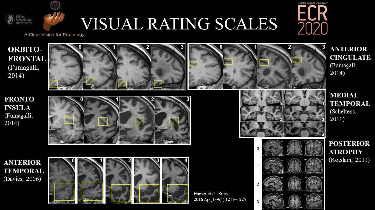
PDF) Evaluation of MR Visual Rating Scales in Major Forms of Dementia

Bringing psychiatrists into the picture: Automated measurement of regional  MRI brain volume in patients with suspected dementia - Pierre Wibawa,  Gabrielle Matta, Sourav Das, Dhamidhu Eratne, Sarah Farrand, Patricia  Desmond, Dennis Velakoulis,
Bringing psychiatrists into the picture: Automated measurement of regional MRI brain volume in patients with suspected dementia - Pierre Wibawa, Gabrielle Matta, Sourav Das, Dhamidhu Eratne, Sarah Farrand, Patricia Desmond, Dennis Velakoulis,

Automated quantitative MRI volumetry reports support diagnostic  interpretation in dementia: a multi-rater, clinical accuracy study |  SpringerLink
Automated quantitative MRI volumetry reports support diagnostic interpretation in dementia: a multi-rater, clinical accuracy study | SpringerLink

Julie Koedam, RHIT - Coding Quality Coordinator/Auditor - BJC HealthCare |  LinkedIn
Julie Koedam, RHIT - Coding Quality Coordinator/Auditor - BJC HealthCare | LinkedIn

Parkinsonism & Related Disorders

Structural imaging findings on non-enhanced computed tomography are  severely underreported in the primary care diagnostic work-up of subjective  cognitive decline | SpringerLink
Structural imaging findings on non-enhanced computed tomography are severely underreported in the primary care diagnostic work-up of subjective cognitive decline | SpringerLink

Transient epileptic amnesia is significantly associated with discrete  CA1-located hippocampal calcifications but not with atrophic changes on  brain imaging - ScienceDirect
Transient epileptic amnesia is significantly associated with discrete CA1-located hippocampal calcifications but not with atrophic changes on brain imaging - ScienceDirect

Visual assessment of posterior atrophy development of a MRI rating scale |  SpringerLink
Visual assessment of posterior atrophy development of a MRI rating scale | SpringerLink

Structural imaging findings on non-enhanced computed tomography are  severely underreported in the primary care diagnostic work-up of subjective  cognitive decline | SpringerLink
Structural imaging findings on non-enhanced computed tomography are severely underreported in the primary care diagnostic work-up of subjective cognitive decline | SpringerLink

Tips for learners of evidence-based medicine: 3. Measures of observer  variability (kappa statistic). - Abstract - Europe PMC
Tips for learners of evidence-based medicine: 3. Measures of observer variability (kappa statistic). - Abstract - Europe PMC

Axial and coronal 3D gradient echo images of a patient with Alzheimer's...  | Download Scientific Diagram
Axial and coronal 3D gradient echo images of a patient with Alzheimer's... | Download Scientific Diagram

PDF) Visual assessment of posterior atrophy development of a MRI rating  scale
PDF) Visual assessment of posterior atrophy development of a MRI rating scale

Analysis of regional atrophy on brain imaging compared with cognitive  function in the elderly and in patients with dementia –
Analysis of regional atrophy on brain imaging compared with cognitive function in the elderly and in patients with dementia –

AVRA: Automatic visual ratings of atrophy from MRI images using recurrent  convolutional neural networks
AVRA: Automatic visual ratings of atrophy from MRI images using recurrent convolutional neural networks

Deep learning for chest radiograph diagnosis: A retrospective comparison of  the CheXNeXt algorithm to practicing radiologists | PLOS Medicine
Deep learning for chest radiograph diagnosis: A retrospective comparison of the CheXNeXt algorithm to practicing radiologists | PLOS Medicine

PDF) Parieto-occipital sulcus widening differentiates posterior cortical  atrophy from typical Alzheimer disease
PDF) Parieto-occipital sulcus widening differentiates posterior cortical atrophy from typical Alzheimer disease

Medial temporal atrophy in preclinical dementia: visual and automated  assessment during six year follow-up
Medial temporal atrophy in preclinical dementia: visual and automated assessment during six year follow-up

Parieto-occipital sulcus widening differentiates posterior cortical atrophy  from typical Alzheimer disease - ScienceDirect
Parieto-occipital sulcus widening differentiates posterior cortical atrophy from typical Alzheimer disease - ScienceDirect

Parieto-occipital sulcus widening differentiates posterior cortical atrophy  from typical Alzheimer disease - ScienceDirect
Parieto-occipital sulcus widening differentiates posterior cortical atrophy from typical Alzheimer disease - ScienceDirect

EPOS™
EPOS™

Voxel-wise deviations from healthy aging for the detection of  region-specific atrophy - ScienceDirect
Voxel-wise deviations from healthy aging for the detection of region-specific atrophy - ScienceDirect
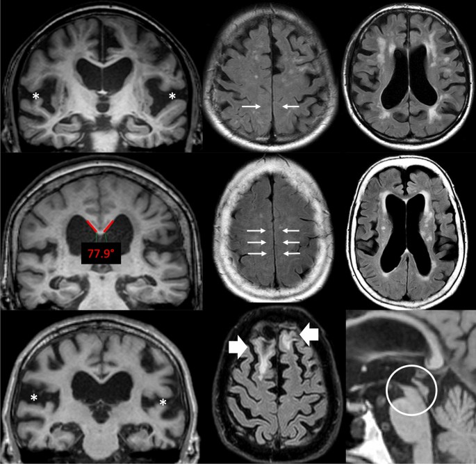
Imaging features associated with idiopathic normal pressure hydrocephalus  have high specificity even when comparing with vascular dementia and  atypical parkinsonism | Fluids and Barriers of the CNS | Full Text
Imaging features associated with idiopathic normal pressure hydrocephalus have high specificity even when comparing with vascular dementia and atypical parkinsonism | Fluids and Barriers of the CNS | Full Text